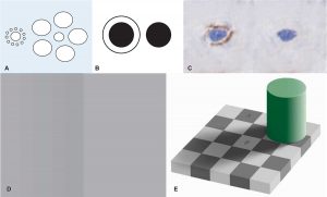
The Gold Standard Paradox in Digital Image Analysis: Manual Versus Automated Scoring as Ground Truth
Abstract CONTEXT: Novel therapeutics often target complex cellular mechanisms. Increasingly, quantitative methods like digital tissue image analysis (tIA) are required to evaluate correspondingly complex biomarkers
WHITE PAPER: The Future of Diagnostics for Immuno-oncology
Executive Summary: The explosion of immuno-oncology (I/O) therapies has created a complicated, dynamic, and competitive landscape that is focused on patient selection for drug efficacy
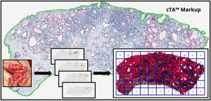
Providing Confidence Around Computational Tissue Analysis Using Heterogeneity Assessments
Abstract: Background: Although the techniques to interrogate the appearance of a biomarker in tissue sections have greatly advanced, there are limitations as to how representative
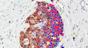
Roles for Pathologists in a High-throughput Image Analysis Team
Abstract Historically, pathologists perform manual evaluation of H&E- or immunohistochemically-stained slides, which can be subjective, inconsistent, and, at best, semiquantitative. As the complexity of staining
Quantitative assessment of pancreatic cancer precursor lesions in IHC-stained tissue with a tissue image analysis platform
Abstract Tissue image analysis (tIA) is emerging as a powerful tool for quantifying biomarker expression and distribution in complex diseases and tissues. Pancreatic ductal adenocarcinoma
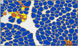
Development of Digital Tissue Image Analysis Solution for Muscle Biopsies in Support of Disease-Modifying Therapies for Duchenne Muscular Dystrophy
Abstract: The continual expression of utrophin protein by pharmacological maintenance of utrophin transcription in dystrophin-deficient muscle fibres is potentially a disease-modifying treatment for Duchenne muscular
