Introduction:
Background
- The modulation of the immune system holds promise in cancer treatment.
- Molecular markers allows for reliable identification and subsequent analysis of individual inflammatory cell types and subtypes to profile the immune component of a tissue.
- Measuring the composition of immune cell types in tumor biopsies can have prognostic value.
- It is thought that similar approaches can predict patient response to a given therapeutic agent.
- Establishing a quantitative paradigms for measuring the composition of immune cell types in tumor biopsies is essential for both prognostic and diagnostic approaches.
- Capturing the quantity and complexity immune infiltration in tumor biopsies requires a whole-tissue based quantitation method capable of integrating multiple inflammatory
cell markers. - In this study, we combined novel advents in tissue image analysis (tIA) to integrate spatial expression of 6 serial-section stained cell-type specific markers in whole tissue clinical
lung cancer samples.
Study Context & Design
- Exploratory proof-of-principle study was designed and engineered to demonstrate full spatial integration of immune cell markers in the context of whole tissues and on cell- by-cell basis.
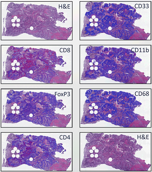
- Serial sections of clinical lung specimens were generated and IHC was performed for CD68, CD4, CD8, CD33, FoxP3, and CD11b markers.
- CellMapTM algorithm was utilized to identify and determine the precise location of individual inflammatory cells in tissues on cell-by-cell basis in the tumor microenvironment
(TME). - Flagship’s FACTS™ (Feature Analysis on Consecutive Tissue Sections) approach was used to integrate six markers (CD68, CD4, CD8, CD33, FoxP3 ,and CD11b )in individual cells onto nascent architecture of the tissue using H&E slides.
- Flagship’s MultivariateMap™ was used to integrate the inflammatory cell types recorded by the 6 markers based on their function and role in immune system biology.
- Using Flagship’s proprietary image analysis tools, we show utility of Flagship’s tIA approaches for pro ling individual inflammatory cell types and providing a comprehensive landscape of the immune system state in tissue biopsies.
- Integrating spatial information of immune cell marker expression will benefit further understanding of cancer pathology and the development of diagnostic tests with clinical value.
