Companion Diagnostic Strategies Specific to Antibody Therapies
Introduction
One premise of antibody-drug conjugates (ADC) is that the bound mAb-antigen complex on the cell surface will internalize and be metabolized by lysosomal proteases to release the free drug. Thus, the efficacy of an ADC is dependent not only on the presence of cell surface antigens, but also on an active system of receptor turnover and receptor-mediated endocytosis.
A predictive assay for patient response would ideally account for both the degree of cell surface expression of the target, as well as cytoplasmic presence of the target to quantify a surrogate for receptor turnover and internalization.
Immunohistochemistry based assays (IHC) in combination with qunantitative tissue image analysis (tIA) are best suited to address these questions as IHC is the only method , which provides the ability to measure both membrane and cytoplasm expression of the target simultaneously within archival FFPE biopsies.
However, the biological mechanisms behind receptor internalization and turnover have not been elucidated for novel therapeutic targets. In most cases, an IHC assay is utilized to evaluate these measures, without prior advance knowledge of how these measures are suitable for patient selection. Unanticipated difficulties in tissue interpretation, such as low apparent expression of the target, occlusion of membrane staining by cytoplasmic staining, or heterogeneity in staining often lead to failure in determining a correct patient stratification approach.
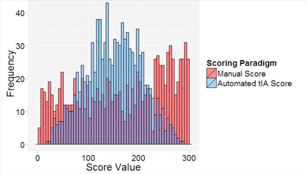
In order to investigate patient selection strategies for ADCs, Flagship Biosciences has invented several proprietary approaches for measuring critical properties of the therapeutic target on the cell surface or inside the cell which can be used to understand and predict efficacy to an ADC using FFPE biopsies.
These tIA based quantitative pathology approaches have been designed to develop a pathology based scoring system for ADC companion diagnostic (CDx) programs and will be useful to:
• Accurately quantify low levels of cell surface target expression;
• Define cell surface target expression independent of cytoplasmic expression;
• Overcome staining heterogeneity; and
• Determine appropriate cut-point(s) based on a target expression for patient selection. These image analysis based approaches can be used to define and evaluate a scoring approach, guide pathologist training, assess objective performance, and best determine a cut-point approach using statistical approaches. These tissue image analysis based tools can be used to create a manual scoring paradigm for an IHC assay or can be incorporated into a medical device directly to support the Premarket Approval (PMA) effort.
